
MRI vs. CT Scan: Understanding the Differences
A radiologist explains what to know about these medical imaging tests, including in which situations each are used and the latest advancements.
Whether it’s to pinpoint the cause of symptoms or a routine screening for a chronic condition, CT and MRIs are critical tools to see what’s going on inside a person’s body. And as improved technology allows for more detailed imaging, radiologists and clinicians can use these scans to better detect what’s happening in bones, muscles, and organs, leading to a more precise diagnosis and the potential for earlier intervention and treatment.
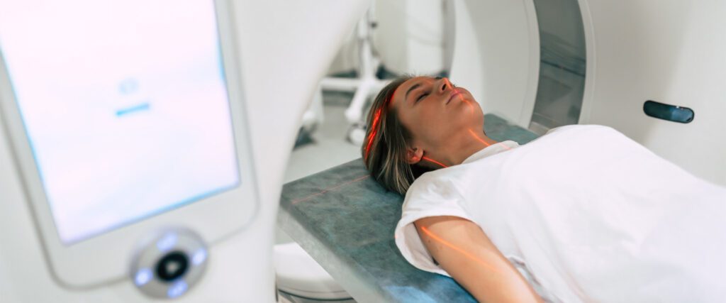
Two of the most common types of advanced imaging are computed tomography (CT) and magnetic resonance imaging (MRI). In both systems the patient lays on a flat surface inside an open tunnel and a detailed three-dimensional image of person’s body is produced. However, they work differently and have different purposes.
Dr. Ajay Gupta, radiologist-in-chief at NewYork-Presbyterian/Columbia University Irving Medical Center, spoke to Health Matters to explain the differences between these imaging systems work, when they are used, and what you should know if you need an MRI or CT scan.
What are the differences between an MRI and a CT scan? When do you need an MRI vs. a CT?
Both MRI and CT are advanced imaging tools that let us look inside the body in great detail, but they work in very different ways. While a traditional X-ray gives you a flat, one-dimensional image, a CT scan uses X-rays to create detailed, cross-sectional images of the body — essentially a 3D X-ray — making it excellent for detecting injuries, lung disease, cancer, and vascular conditions.
MRI, on the other hand, uses powerful magnets and radio waves rather than radiation. It gives us very precise views of soft tissues like the brain, spine, joints, and organs. The choice between them depends on what we’re looking for. For example, CT tends to be faster and better for emergencies, while MRI offers more detail for certain tissues and conditions.
What are some advancements in specialized imaging, like photon-counting CT scans?

Photon-counting CT is the most significant leap forward in CT technology in decades. Traditional CT scanners measure the overall energy from X-rays passing through the body. Photon-counting CT, however, counts individual X-ray photons and measures their energy precisely. This gives us much sharper images, allows us to see smaller structures, and can even help differentiate between materials like calcium, iodine, or soft tissue more accurately — all with less radiation dose.
At NewYork-Presbyterian, we’re proud to have the first photon-counting CT scanner across our entire system, located at NewYork-Presbyterian The One in Westchester. It’s truly a game changer. For instance, we can now see the composition of subtle coronary artery plaques in the heart in ways never before possible. By seeing the earliest manifestation of coronary artery disease on a CT scan before a patient has any symptoms, we equip our doctors and patients with the critical information they need to help reduce the risk of a future heart attack. Another example is for the evaluation of hearing loss, where the exceptional spatial resolution of our new scanners lets us visualize tiny structures — like the ossicles in the inner ear — that were previously difficult to assess. This technology gives us clearer, faster, and safer answers for our patients.
What are some circumstances where someone would need a specialized MRI or CT scan?
No two patients are exactly alike, and we are focused on tailoring our imaging exams to meet the need of every patient who comes to us for their care. Specialized imaging comes into play when we need greater precision or are looking at a very specific part of the body. For example, cardiac CT or MRI can give detailed pictures of the heart and vessels; advanced MRI techniques can assess brain function or detect early signs of neurodegenerative disease; and specialized CT can characterize lung nodules or detect subtle bone injuries. Customizing the scan to the patient and the question at hand allows us to get the most meaningful results.
What is the difference in radiation exposure between MRI and CT scans?
MRI does not use radiation at all, whereas CT uses radiation. That said, modern CT scanners — including photon-counting CT — use dramatically lower doses of radiation than older generations did, thanks to newer hardware in the scanners, such as advanced detectors. The benefits of an accurate diagnosis almost always outweigh the small potential risks of radiation exposure.
For people who need regular imaging, are there any radiation or health concerns?
For most patients, the risk from radiation exposure is extremely low, especially with the dose reductions we’ve achieved in recent years. When someone needs frequent imaging — say, for chronic conditions or cancer follow-up — we carefully track exposure and use MRI or other alternatives, like ultrasound technology, which uses high-frequency sound waves to create images.
What is dye contrast and when is that used in imaging?
Contrast agents are special dyes that enhance what we can see on scans. In CT, they help highlight blood vessels and organs. In MRI, contrast improves our ability to detect inflammation, tumors, or blood-brain barrier changes. They’re safe for most people, and we screen patients carefully to ensure appropriate use.
Do you always have to lay perfectly still when getting an MRI or CT scan?
For the most part, to capture high-quality images, it’s helpful if our patients remain as still as possible. There are things we can’t control that we can capture, like a beating heart, but motion can degrade the quality of the image a little bit.
That said, not all imaging involves laying down. At Och Spine at NewYork-Presbyterian/The Spiral, we can look at a person’s whole skeletal system while standing in a natural position. This can be helpful to diagnose conditions like scoliosis that cause the spine to curve. For this, we can use a specialized imaging tool called EOS imaging. This new technology uses very low doses of radiation to capture 3-D pictures of the skeleton from multiple angles in one set of images, all while a person is standing up.
What do you tell people who are nervous about getting a scan, whether about radiation, contrast, or claustrophobia?
First, we reassure patients that these procedures are extremely safe and that their comfort and well-being are our top priorities. We walk them through what to expect, answer any questions they may have, use calming techniques, and when appropriate, mild medication to help with anxiety. We have blankets if the room gets cold, earplugs because the machines are loud, and we can play calming music through headphones.
Our technologists are excellent at helping patients manage claustrophobia through compassion, communication, and reassurance.
What’s the future of imaging technology?
Medical imaging accounts for an enormous share of the data we generate from patient encounters in health care. I’m excited about how artificial intelligence will allow us to make more precise and accurate diagnoses and at the earliest stage possible when treatment is most effective.
New technologies are also enabling us to acquire medical images much faster than before. MRI scans that used to take 30 minutes or longer can be done in 10 minutes or less using the newer technologies — including AI based tools — that are being built into new MRI scanners. These advances make each patient’s experience much more comfortable and efficient.