
Understanding Breast Cancer Screening
A breast radiologist shares how mammograms, ultrasounds and MRIs are used to help detect and prevent breast cancer.
Breast cancer is the second most common cancer among all women in the United States, with an estimated 300,000 new cases in 2024, according to the American Cancer Society (ACS). But the good news is that we have more tools than ever to help screen for breast cancer. “Cases that we find early are treatable,” says Dr. Katja Pinker-Domenig, chief of the Division of Breast Imaging at NewYork-Presbyterian/Columbia University Irving Medical Center. “If somebody has a diagnosis of breast cancer, the sooner we can address the problem, the better.”

Dr. Katja Pinker-Domenig
In April 2024, the U.S. Preventive Services Task Force updated its screening guidelines, recommending that women who are at average risk for breast cancer begin breast cancer screening at age 40. Also, the U.S. Food and Drug Administration (FDA) now requires mammogram centers in every state to notify patients if they have dense breast tissue. About half the women in the U.S. have high breast density, a risk factor for breast cancer that often requires supplemental testing.
“We are going into a more layered way of screening, tailoring it to the risk,” says Dr. Pinker-Domenig. “Not everybody who has extremely dense breast tissue has the same breast cancer risk as somebody who has, for instance, a strong family history, which would put them in another risk level.”
Health Matters spoke with Dr. Pinker-Domenig about the different screening methods for breast cancer, including mammograms, ultrasounds, and MRIs, and when these different methods are recommended.
In the U.S., mammograms are the gold standard for breast cancer screening. What can they detect?
Dr. Pinker-Domenig: Women at average risk for breast cancer are recommended to start with mammograms, which are a low-dose x-ray that looks at breast tissue. The machine has two plates that compress the breast to spread tissue apart before a picture is taken.
In a mammogram, we are not only looking for masses but also details like asymmetries and microcalcifications that can indicate that something might be growing at that spot.
There are different types of mammograms. In a regular mammogram, which is 2D, there can be overlapping tissue, and patients may get recalled because we are not sure if it is something of concern. Digital breast tomosynthesis, sometimes referred to as a 3D mammogram, is another type of mammogram that allows us to both lower recall rates and find more cancers.
The newest development is contrast-enhanced mammography, where we use a dye injected into the veins to help visualize cancers. It is a great option in women with greater than average risk and who have high breast density, where mammography or even tomosynthesis are limited. So, it is an exciting new addition to the screening toolbox that we have.
It’s also important to note that in the screening process, self-breast exams are still valuable because you know your body. If you feel there is something different, I recommend having it checked.
When do people need supplemental screening, such as an ultrasound?
If there is a finding detected on a mammogram, a diagnostic breast ultrasound is often part of the work up. However, ultrasound can also be used for supplemental screening in women with dense breasts.
While ultrasound is helpful in finding more cancers, it can also generate more false positives. I sometimes get the question: Can I have an ultrasound instead of a mammogram? The answer is no because in ultrasounds, we cannot see microcalcifications, which are often the earliest form of breast cancer. An ultrasound alone is not an appropriate screening test.
Who is recommended to get an MRI and how are they used for screening?
An MRI is undoubtedly the most sensitive test for breast cancer detection, regardless of risk factors, such as breast density. However, MRI is also the most expensive test. It is currently recommended for patients at high risk for breast cancer, such as those who have a 20% lifetime risk or more. The standard screening for these high-risk women is an MRI plus a mammography because MRIs can also miss the early small cancers that we see as microcalcifications on the mammogram. Ultrasounds are not needed for patients getting a mammogram and MRI.
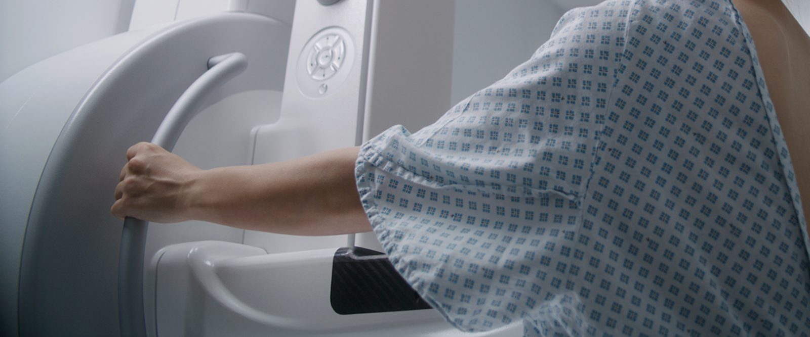
How does a breast MRI work?
A breast MRI is a machine that uses strong magnets and radio waves to create detailed pictures of the inside of your breast. Like in contrast enhanced mammography, in breast MRI we use a dye injected into the veins to highlight areas where a cancer is growing. Unlike mammograms, MRI doesn’t use radiation and is completely painless. It involves lying still inside the scanner that looks like a tunnel. However, if you are claustrophobic and do not like closed spaces you may feel a little uncomfortable being inside.
In what scenarios would doctors do a biopsy?
A scenario where a biopsy is recommended is if there is anything suspicious for malignancy. I usually advise patients to not immediately think you have a cancer. We’re recommending a biopsy if the likelihood of malignancy is greater than 2% because we do not want to overlook something.
The initial biopsy is not a surgery. The radiologist uses imaging guidance and local anesthesia to take a small tissue sample to study it. During the biopsy, you will feel the radiologist doing something, but it is not painful. I personally compare it to the dentist.
When we are done with the biopsy, we put a tiny clip marker at that area where we took the sample. This is important because if that comes back as benign, it will show whoever reads future mammograms that a biopsy has been done and that results were benign. In case you need surgery, this also helps with knowing exactly where to go, and the surgeon can be as tissue-sparing as possible. The marker doesn’t set off metal detectors, such as at the airport, and it is non-allergic.
What should people keep in mind about screening if they are going into it for the first time, or in general?
If it is the first time you have ever had a screening exam, if you get recalled, do not immediately panic. Since we do not have an old image for comparison, we want to look at everything and document everything properly. So, if on a baseline exam you happen to have a recall, that doesn’t necessarily mean that you have cancer.
Patients should also be aware that we want to err on a side of caution and make sure that we catch cancers as early as possible. Almost everything that we find early nowadays is treatable. That is why it is so important to have your screening mammogram.
Katja Pinker-Domenig, M.D., is an attending radiologist at NewYork-Presbyterian/Columbia University Irving Medical Center. She is also a professor of radiology at Columbia University Vagelos College of Physicians and Surgeons and chief of the Division of Breast Imaging.
Dr. Pinker-Domenig, an internationally renowned physician-scientist and board-certified radiologist with expertise in breast imaging, has extensive expertise in all aspects of state-of-the-art breast imaging and interventions, including mammography, digital breast tomosynthesis, ultrasound, MRI, and biopsy procedures.
Additional Resources
Learn more about breast cancer screening at NewYork-Presbyterian.